
High-yield map
Key points before the detail
- 01
Start with physiology and time-critical syndromes before naming a disease.
- 02
Give analgesia early; appropriate analgesia does not prevent accurate assessment.
- 03
Pain mechanism and migration often carry more information than a pain adjective alone.
- 04
Choose imaging to answer a specific question: perfusion, perforation, obstruction, bleeding, biliary or pelvic pathology.
- 05
A normal early lactate, white-cell count or CRP does not exclude serious intra-abdominal pathology.
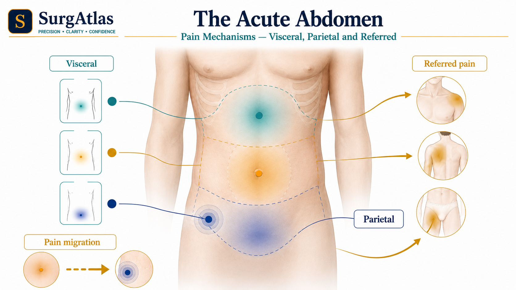
1. Ask the dangerous question first
Undifferentiated abdominal pain is a syndrome, not a diagnosis. Before constructing a long differential, identify whether the patient has shock, generalised peritonism, bowel ischaemia, perforation, strangulation, uncontrolled bleeding or sepsis. These findings determine urgency and often change the order of investigation and treatment.
Diagnosis and resuscitation should proceed together. A deteriorating patient should not wait for diagnostic certainty before senior surgical, anaesthetic or critical-care involvement.
2. Use pain mechanism to organise anatomy
Migration is especially useful because it may reflect changing anatomy. Classic appendicitis begins as vague periumbilical visceral pain and localises to the right iliac fossa as adjacent parietal peritoneum becomes inflamed.
| Pain mechanism | Typical quality | Anatomical meaning |
|---|---|---|
| Visceral | Dull, poorly localised, often midline | Stretch, distension or ischaemia of a viscus; foregut tends to epigastrium, midgut periumbilical region and hindgut lower abdomen. |
| Parietal | Sharp, well localised, worse with movement or coughing | Irritation of parietal peritoneum; guarding and percussion tenderness may develop. |
| Referred | Pain felt away from the diseased organ | Convergence of visceral and somatic afferents; for example diaphragmatic irritation may refer to the shoulder. |
3. The first ten minutes: A–E plus immediate actions
- Keep nil by mouth until aspiration risk and need for urgent procedure are clear.
- Give analgesia and antiemetics early, then reassess.
- Give prompt antibiotics when intra-abdominal sepsis, perforation, cholangitis or another bacterial emergency is suspected, following local antimicrobial policy.
- Escalate early for instability, peritonism, suspected ischaemia/strangulation, free perforation, uncontrolled bleeding or high-risk diagnostic uncertainty.
| Step | Key actions |
|---|---|
| Airway | Confirm patency; seek early anaesthetic help if consciousness is reduced or major haematemesis threatens airway protection. |
| Breathing | Respiratory rate, oxygen saturation and chest examination; remember thoracic disease can present as abdominal pain. |
| Circulation | Pulse, blood pressure, capillary refill and perfusion; IV access, bloods and reassessed fluid resuscitation when hypovolaemic. |
| Disability | Mental state, glucose and early analgesia. |
| Exposure | Inspect the abdomen, groins, scars, stomas, back and skin while preserving dignity and temperature. |
4. History and examination that change probability
- Pain: site, migration, onset, tempo, colicky versus constant character, radiation, provoking factors and trajectory.
- Associated symptoms: vomiting, last stool/flatus, diarrhoea or bleeding, urinary symptoms, pregnancy possibility and gynaecological symptoms, fever, rigors and syncope.
- Background: previous abdominal or bariatric surgery, known hernia, gallstones, IBD, ulcer disease, malignancy, vascular disease and medication such as NSAIDs, steroids, antithrombotics or opioids.
- Examination: look before touching, palpate gently away from pain, assess involuntary guarding/percussion tenderness, examine relevant hernial orifices and complete chest/cardiovascular examination.
- DRE, pelvic and testicular examinations are selective: perform them when the result answers a clinical question, with consent and a chaperone.
5. Red flags that should change your tempo
- Shock, syncope, active GI bleeding or a rapidly falling haemoglobin.
- Generalised guarding/rigidity, marked percussion tenderness or evidence of free perforation.
- Pain out of proportion to examination, metabolic acidosis or embolic/atherosclerotic risk: consider mesenteric ischaemia.
- Persistent severe pain in obstruction, focal tenderness, fever, tachycardia, rising lactate or a closed-loop CT pattern: consider strangulation/ischaemia.
- Painful irreducible hernia, skin discolouration or systemic toxicity.
- Pregnancy with pain, bleeding, syncope or shoulder-tip pain: exclude ectopic pregnancy.
- Previous bariatric surgery with severe or intermittent unexplained pain: internal hernia can have deceptively mild findings.
- Immunosuppression or corticosteroid use may blunt fever and peritonism.
6. Investigations: ask what the test must answer
| Investigation | Use it to answer |
|---|---|
| Bedside | Physiological trend, glucose, urine findings, cardiac mimics/risk and pregnancy status where relevant. |
| Bloods | Anaemia, inflammation, renal/electrolyte disturbance, hepatobiliary or pancreatic clues, coagulation and perfusion/lactate in the unwell patient. |
| Ultrasound | Biliary disease, pelvic/early pregnancy questions and selected hernias or younger-patient pathways. |
| CT abdomen/pelvis with IV contrast | Perforation, obstruction, diverticulitis, abscess, complicated appendicitis, malignancy and many adult acute-abdomen questions. |
| CT angiography | Mesenteric ischaemia or ongoing major GI bleeding where arterial localisation matters. |
| CT KUB | Typical renal colic when imaging is required; not a generic acute-abdomen scan. |
7. A five-step MRCS viva structure
This structure keeps an answer safe when the exact diagnosis is uncertain. It also prevents a common viva failure: reciting a differential while ignoring deteriorating physiology.
- 1. State the dominant syndrome and immediate threats.
- 2. Give the first resuscitation actions.
- 3. Name two or three discriminating history/examination findings.
- 4. Choose the first-line investigation and explain what it must answer.
- 5. State the escalation threshold and likely source-control route.
Active recall
Close the notes and answer these
Try each question from memory before revealing the answer. These public prompts are a small preview of the integrated retrieval system inside SurgAtlas.
01What should be your first question in an undifferentiated acute abdomen?
Whether the patient is physiologically unstable or has evidence of a time-critical syndrome such as peritonitis, bowel ischaemia, perforation, strangulation, major haemorrhage or sepsis.
02How do visceral and parietal pain differ?
Visceral pain is typically dull and poorly localised from organ stretch/distension/ischaemia; parietal pain is sharper and well localised because the parietal peritoneum is somatically innervated.
03What abdominal finding should never be omitted when assessing possible obstruction?
Examination of relevant hernial orifices.
04When is CT angiography particularly useful?
When mesenteric ischaemia or ongoing major GI bleeding is suspected and arterial perfusion/localisation must be assessed.
05Can a normal early lactate exclude mesenteric ischaemia?
No. A normal early lactate does not exclude bowel ischaemia; clinical context, imaging and repeated assessment are essential.
Sources & editorial basis
References & editorial basis
- SurgAtlas production source. General Surgery — The Acute Abdomen, clinical and publication review completed 17 July 2026.
- NICE. Related emergency management principles should be applied alongside current condition-specific NICE guidance and local pathways; this lesson is educational and not a substitute for local acute-care protocols. Source ↗
This lesson is derived from the corresponding SurgAtlas production teaching material. Where the source makes current management or guideline claims, the public lesson uses the cited contemporary guidance. It is written for education and examination preparation, not as patient-specific clinical advice.
Editorial details
Medical Doctor (MD) · MRCS Part A · Physician · Surgical Educator
SurgAtlas is an educational resource. For patient care, verify current national guidance, local antimicrobial and transfusion policies, specialty pathways and individual patient factors.
Continue in SurgAtlas
Turn reading into retrieval.
Use the full learning workspace for chapter progress, integrated questions, review scheduling and exam-focused study tools. Free accounts do not include recurring AI usage.