
High-yield map
Key points before the detail
- 01
Classify by level, completeness, blood supply, geometry and mechanism.
- 02
CT with IV contrast is usually the key adult investigation for transition point, cause and threatened bowel.
- 03
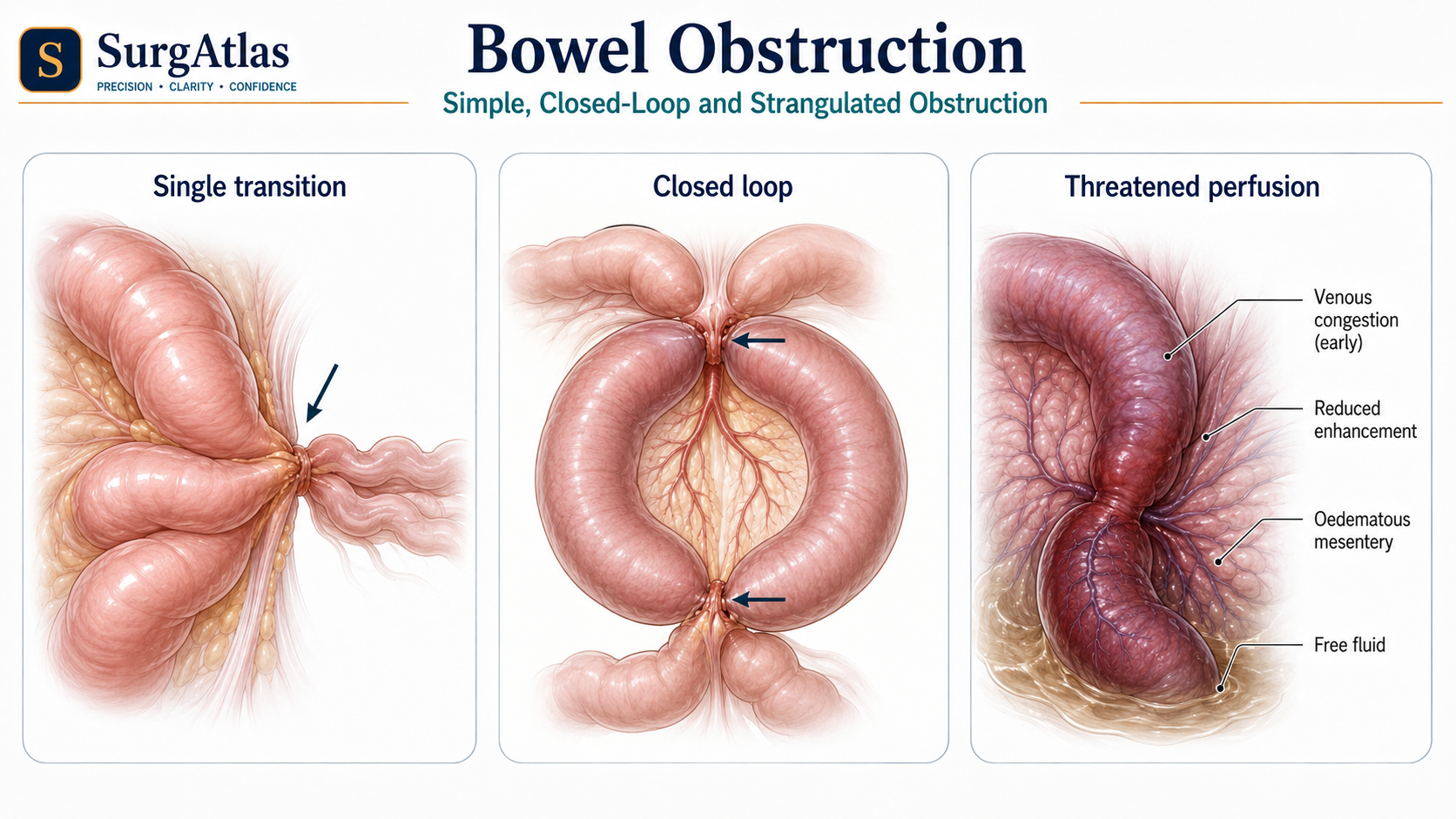
A closed loop is obstructed at two points and may become ischaemic rapidly.
- 04
Nasogastric decompression is selective, not a ritual for every mild partial obstruction.
- 05
The “72-hour” adhesive-SBO period is an upper boundary for selected stable patients, never permission to wait through deterioration.
1. Classify before memorising causes
| Axis | Categories | Why it matters |
|---|---|---|
| Level | Small bowel vs large bowel | Changes likely causes, vomiting pattern, distension and definitive treatment. |
| Completeness | Partial vs complete | Partial obstruction is more likely to resolve non-operatively; complete obstruction has a higher failure rate. |
| Blood supply | Simple vs strangulated | Strangulation is a surgical emergency because impaired venous outflow can progress to arterial compromise and necrosis. |
| Geometry | Single transition vs closed loop | A closed loop is blocked at two points and can distend rapidly. |
| Mechanism | Mechanical vs ileus/pseudo-obstruction | Mechanical obstruction needs a transition/cause; functional obstruction requires treatment of the precipitant. |
2. Causes and clinical pattern
Postoperative adhesions are a common cause of small-bowel obstruction. Other important causes include incarcerated hernia, Crohn stricture, tumour, volvulus, intussusception and gallstone ileus. In large-bowel obstruction, colorectal cancer and volvulus are high-yield causes alongside diverticular stricture and faecal impaction.
Colicky pain reflects peristaltic contraction against a blockage. Vomiting tends to occur earlier in proximal small-bowel obstruction; distension is often greater in distal small-bowel and large-bowel obstruction. Obstipation supports complete obstruction, but early stool passage may simply empty bowel distal to the block.
3. Recognise threatened bowel
- Pain becoming continuous, severe or focal rather than purely colicky.
- Focal tenderness, guarding or peritonism.
- Fever, persistent tachycardia or worsening physiological instability.
- Metabolic acidosis or rising lactate — but a normal lactate does not exclude early ischaemia.
- CT evidence of a closed loop, poor bowel-wall enhancement, free fluid in the appropriate context or perforation.
4. Examination: do not forget the hernias
- A–E assessment and hydration/perfusion status with serial observations.
- Inspect for scars, distension, visible peristalsis and stomas.
- Examine every relevant hernial orifice.
- Palpate gently for focal tenderness, masses and peritonism.
- Do not rely on bowel sounds to rule obstruction in or out.
- Use digital rectal examination selectively when faecal impaction, rectal tumour, blood or another finding would change immediate management.
- If ileus is possible, actively seek sepsis, pancreatitis, electrolyte disturbance, medication effects and postoperative complications.
5. CT should answer four questions
CT abdomen/pelvis with IV contrast is usually the key adult investigation because it can answer all four questions. Plain abdominal radiography may show dilated bowel or a volvulus pattern, but it is less accurate for cause and complications and should not delay CT when concern persists.
In selected adhesive small-bowel obstruction without peritonitis, strangulation or ischaemia, a water-soluble contrast challenge can support decision-making. Passage of contrast into the colon within 24 hours predicts likely non-operative resolution.
- Is there an obstruction and where is the transition point?
- What is the cause?
- Is the geometry simple or closed loop?
- Is the bowel threatened — poor perfusion, ischaemia or perforation?
6. Immediate management
- Nil by mouth, IV access, analgesia and antiemetics.
- Correct sodium, chloride, potassium and water deficits with reassessed fluid therapy.
- Use nasogastric decompression for persistent vomiting, marked gastric/small-bowel distension, aspiration risk or as part of a contrast pathway; it is not mandatory for every mildly symptomatic partial obstruction.
- Use urinary catheterisation when shock, major replacement or precise urine-output monitoring justifies it.
- Give antibiotics when strangulation, perforation, sepsis or operative contamination is suspected, following local policy.
- Provide VTE prophylaxis unless contraindicated and review nutrition if obstruction is prolonged.
7. Adhesive small-bowel obstruction: when non-operative care is appropriate
| Suitable monitored pathway | Escalate to operation |
|---|---|
| Probable adhesions, stable physiology, no peritonitis, strangulation, ischaemia or perforation. | Peritonitis, deterioration, incarcerated hernia, perforation, CT devascularisation/closed loop or unresolved diagnostic concern. |
| Resuscitation, bowel rest, selective NG decompression, serial examination/labs and water-soluble contrast according to local protocol. | Urgent adhesiolysis and assessment of bowel viability; resect non-viable bowel when required. |
| A closely monitored trial can be appropriate while objective progress is occurring. | Failure to resolve by about 72 hours should prompt reassessment for surgery; operate earlier whenever danger signs appear. |
Active recall
Close the notes and answer these
Try each question from memory before revealing the answer. These public prompts are a small preview of the integrated retrieval system inside SurgAtlas.
01What five axes should you use to classify bowel obstruction?
Level, completeness, blood supply, geometry and mechanism.
02Why is closed-loop obstruction dangerous?
The segment is blocked at two points, so pressure can rise rapidly and compromise venous then arterial blood flow, leading to ischaemia, necrosis and perforation.
03What must CT establish in suspected bowel obstruction?
Presence/level of obstruction, transition point and cause, closed-loop geometry and whether bowel perfusion or integrity is threatened.
04Does a normal lactate exclude strangulation?
No. Early bowel ischaemia may occur with a normal lactate; the whole clinical and CT picture matters.
05When is non-operative management inappropriate in adhesive SBO?
When there is peritonitis, strangulation/ischaemia, perforation, deterioration, an incarcerated hernia, a dangerous CT pattern or another unresolved concern requiring surgery.
Sources & editorial basis
References & editorial basis
- SurgAtlas production source. General Surgery — Bowel Obstruction: mechanism-based pathway from decompression and CT to timely surgery.
- World Society of Emergency Surgery. Bologna guidelines for diagnosis and management of adhesive small bowel obstruction: 2017 update. Source ↗
This lesson is derived from the corresponding SurgAtlas production teaching material. Where the source makes current management or guideline claims, the public lesson uses the cited contemporary guidance. It is written for education and examination preparation, not as patient-specific clinical advice.
Editorial details
Medical Doctor (MD) · MRCS Part A · Physician · Surgical Educator
SurgAtlas is an educational resource. For patient care, verify current national guidance, local antimicrobial and transfusion policies, specialty pathways and individual patient factors.
Continue in SurgAtlas
Turn reading into retrieval.
Use the full learning workspace for chapter progress, integrated questions, review scheduling and exam-focused study tools. Free accounts do not include recurring AI usage.