
High-yield map
Key points before the detail
- 01
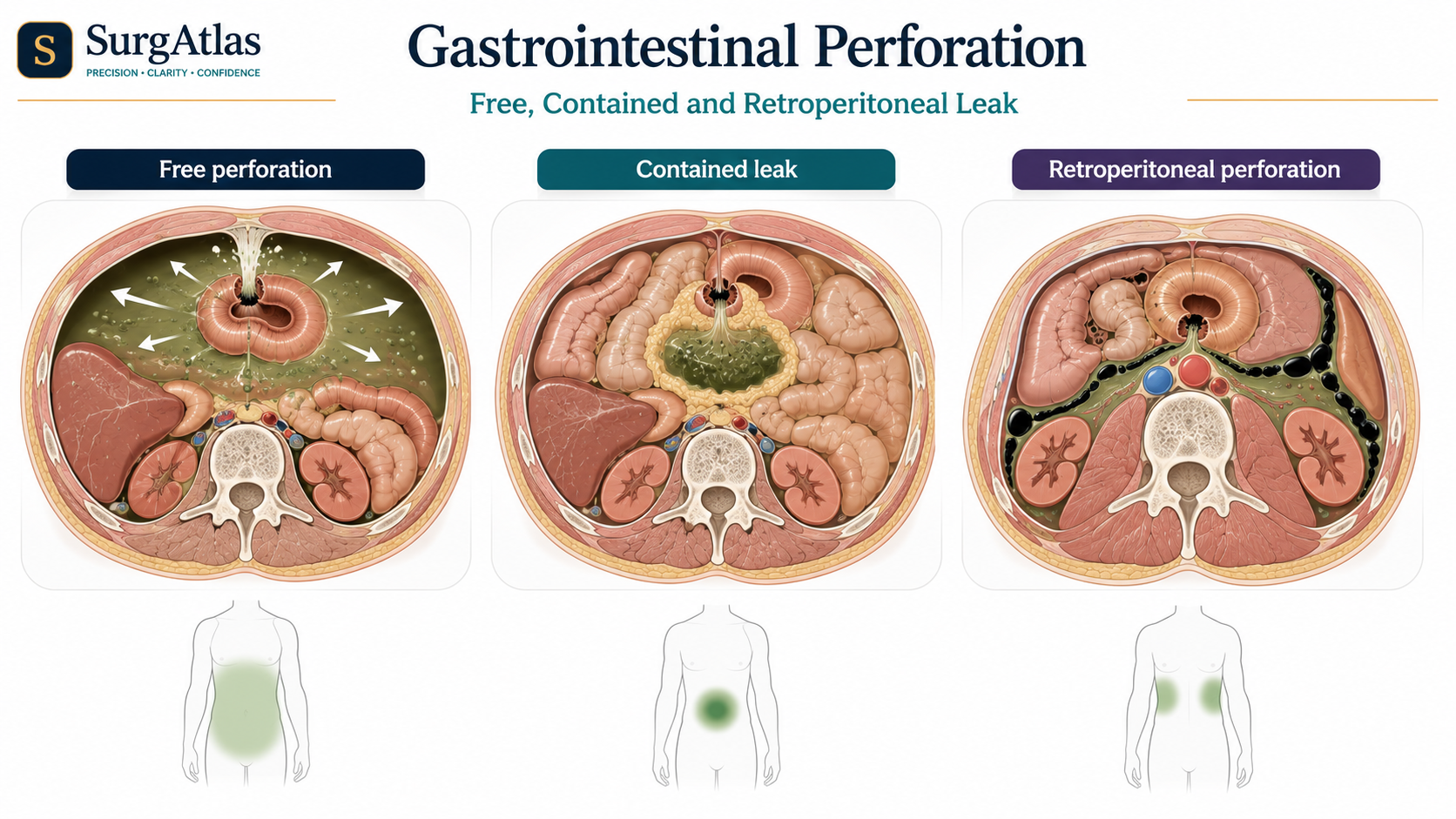
Perforation may be free, contained or retroperitoneal.
- 02
CT with IV contrast is the key investigation in most stable adults.
- 03
A normal plain film does not exclude perforation.
- 04
Antibiotics support the patient; source control treats the cause.
- 05
Anastomosis versus stoma depends on physiology, contamination and tissue quality.
1. Why perforation is dangerous
A perforation allows gas, fluid and microorganisms to escape beyond the bowel lumen. A small sealed leak may cause local inflammation, while free colonic perforation can rapidly produce faecal peritonitis, shock and multiorgan failure.
2. Free, contained and retroperitoneal leaks
| Pattern | Clinical meaning |
|---|---|
| Free perforation | Wide peritoneal contamination with generalised guarding, sepsis and a high likelihood of operative source control. |
| Contained perforation | Omentum or adjacent tissues limit spread; selected stable patients may be managed with antibiotics, drainage and reassessment. |
| Retroperitoneal perforation | Back or flank pain and retroperitoneal gas may occur with little obvious intraperitoneal free air. |
3. Immediate assessment and resuscitation
- A–E assessment with early senior surgical and anaesthetic review when unwell.
- IV access; FBC, U&E, LFT, CRP, coagulation, group-and-save/crossmatch and blood gas with lactate according to severity.
- Keep nil by mouth, give analgesia and antiemetic therapy, and use reassessed crystalloid boluses for hypovolaemia.
- Start broad-spectrum IV antibiotics promptly when contamination is suspected, following local antimicrobial policy.
- Use nasogastric decompression selectively for vomiting, upper-GI perforation, gastric distension or obstruction.
4. Imaging answers the source-control question
| Test | Role |
|---|---|
| CT abdomen/pelvis with IV contrast | Preferred in most stable adults; identifies site, gas/fluid, abscess, tumour, obstruction, ischaemia and a drainage route. |
| Erect chest radiograph | May show subdiaphragmatic free gas when CT is not immediately available, but a normal film does not exclude perforation. |
| Ultrasound | Useful in selected children, pregnancy and focal pathology, but does not replace CT in a deteriorating adult. |
5. Source-control decision
| Situation | Likely approach |
|---|---|
| Diffuse peritonitis, shock, ongoing leak or non-viable bowel | Urgent operation with repair/resection, washout and assessment of viability. |
| Localised abscess in a stable patient | Antibiotics with image-guided drainage when anatomically feasible. |
| Selected sealed upper-GI leak | Closely monitored non-operative care only when stable, without sepsis/peritonitis and with immediate surgical capability. |
| Profound physiological derangement | Damage-control surgery may prioritise rapid contamination control over definitive reconstruction. |
6. Operative principles
- Control contamination by closing, resecting or diverting the leaking segment.
- Remove non-viable tissue and inspect the bowel when ischaemia or closed-loop obstruction is possible.
- Choose primary anastomosis only when physiology, tissue, contamination and operative conditions are acceptable.
- Take appropriate specimens from suspicious lesions without delaying lifesaving source control.
Active recall
Close the notes and answer these
Try each question from memory before revealing the answer. These public prompts are a small preview of the integrated retrieval system inside SurgAtlas.
01What is the key danger in gastrointestinal perforation?
Contamination leading to peritonitis, sepsis and physiological deterioration, especially when source control is delayed.
02Does a normal erect chest radiograph exclude perforation?
No. Contained and retroperitoneal leaks may have no obvious free intraperitoneal air.
03What is the preferred imaging test in most stable adults?
Contrast-enhanced CT of the abdomen and pelvis.
04When is non-operative treatment of a perforation reasonable?
Only in carefully selected stable patients with a contained or proven sealed leak, no diffuse peritonitis/sepsis, reliable monitoring and immediate access to surgery.
05What determines primary anastomosis versus stoma?
Patient physiology, contamination, bowel viability/tissue quality and operative conditions.
Sources & editorial basis
References & editorial basis
- SurgAtlas production source. General Surgery — Gastrointestinal Perforation. Public lesson curated from the corresponding production chapter.
- RCS England / emergency general surgery standards. Emergency source-control principles should be applied with current local sepsis, antimicrobial and imaging pathways.
This lesson is derived from the corresponding SurgAtlas production teaching material. Where the source makes current management or guideline claims, the public lesson uses the cited contemporary guidance. It is written for education and examination preparation, not as patient-specific clinical advice.
Editorial details
Medical Doctor (MD) · MRCS Part A · Physician · Surgical Educator
SurgAtlas is an educational resource. For patient care, verify current national guidance, local antimicrobial and transfusion policies, specialty pathways and individual patient factors.
Continue in SurgAtlas
Turn reading into retrieval.
Use the full learning workspace for chapter progress, integrated questions, review scheduling and exam-focused study tools. Free accounts do not include recurring AI usage.