
High-yield map
Key points before the detail
- 01
A normal or near-normal blood pressure does not exclude compensated shock.
- 02
Use the whole perfusion picture: mental state, capillary refill, skin temperature, pulse, urine output, lactate/base deficit and trend.
- 03
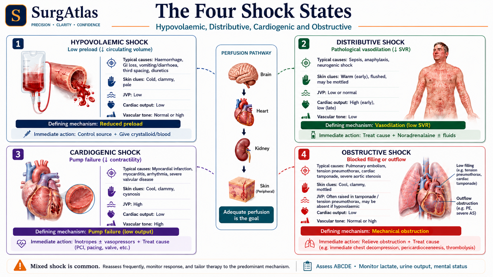
The four broad mechanisms are hypovolaemic, distributive, cardiogenic and obstructive; mixed physiology is common.
- 04
In active major haemorrhage, activate the local major haemorrhage pathway early and prioritise haemorrhage control and blood-component resuscitation rather than repeated large crystalloid volumes.
Start with the physiology
Shock develops when the circulation cannot deliver enough oxygen to meet cellular requirements. Oxygen delivery depends on cardiac output and arterial oxygen content. A patient can therefore become shocked because there is too little circulating volume, inadequate pump function, profoundly abnormal vascular tone, mechanical obstruction to filling/outflow, insufficient oxygen-carrying capacity, or a combination.
Four physiological patterns
| Type | Typical physiology | Useful clues |
|---|---|---|
| Hypovolaemic | Reduced preload and cardiac output with compensatory vasoconstriction | Haemorrhage or fluid loss, cool peripheries, narrow pulse pressure, low filling |
| Distributive | Low systemic vascular resistance; output may be high early | Sepsis, anaphylaxis or neurogenic causes; warm vasodilated pattern can occur |
| Cardiogenic | Pump failure with low output and raised filling pressures | Pulmonary oedema, raised JVP, chest pain, arrhythmia or new murmur |
| Obstructive | Mechanical restriction to cardiac filling or outflow | Tension pneumothorax, tamponade or massive pulmonary vascular obstruction patterns |
Recognise shock before hypotension
- New confusion, restlessness or collapse.
- Tachycardia or an unexpectedly rising pulse for the patient.
- Cool, clammy or mottled skin with delayed capillary refill; septic shock can be warm early.
- Falling urine output and worsening renal function.
- Rising lactate or worsening base deficit/acidaemia in the appropriate context.
- Increasing oxygen requirement or tachypnoea as compensation and organ dysfunction develop.
Immediate priorities
| Step | Actions |
|---|---|
| A/B | Call for help; protect the airway; give oxygen to the appropriate target; look immediately for tension pneumothorax, major haemothorax and other reversible chest threats. |
| C | Monitoring, two short wide-bore IV cannulae where possible, blood sampling/crossmatch, external haemorrhage control, warm the patient and initiate the appropriate resuscitation pathway. |
| D/E | Assess consciousness/glucose; fully expose while preventing heat loss; inspect wounds, drains, abdomen, pelvis, long bones and operative sites. |
| Definitive control | Operating theatre, endovascular/interventional radiology, endoscopy or other cause-specific treatment according to the source and physiological response. |
Major haemorrhage: principles rather than a universal pack
Major-haemorrhage pack composition, emergency group-O policy, component ratios outside specific trauma pathways, calcium replacement and anticoagulant reversal protocols vary between organisations. Public teaching should therefore emphasise early protocol activation, balanced blood-component resuscitation guided by clinical/laboratory information, prevention of hypothermia, correction of coagulopathy and rapid haemorrhage control rather than inventing one universal regimen.
- In active bleeding, haemoglobin can initially appear deceptively normal because plasma and red cells are lost together.
- A transient response to resuscitation suggests ongoing loss until proved otherwise.
- Repeated investigations must not delay definitive control in a patient who remains unstable.
Active recall
Close the notes and answer these
Try each question from memory before revealing the answer. These public prompts are a small preview of the integrated retrieval system inside SurgAtlas.
01Why can a patient be in shock with a normal blood pressure?
Compensatory vasoconstriction and tachycardia can preserve central pressure while organ and peripheral perfusion are already failing.
02What are the four broad physiological types of shock?
Hypovolaemic, distributive, cardiogenic and obstructive.
03What is the central management principle in haemorrhagic shock?
Resuscitate while rapidly identifying and definitively controlling the bleeding source; do not allow repeated fluid or investigations to postpone control.
Sources & editorial basis
References & editorial basis
- SurgAtlas production chapter — Shock and Major Haemorrhage. Primary SurgAtlas source. Guideline-dependent statements in this public lesson were separately checked against the current authoritative sources listed below.
- NICE — Major trauma: assessment and initial management (NG39). Authoritative UK framework for major-trauma assessment and haemorrhage-control principles. Source ↗
This lesson is derived from the corresponding SurgAtlas production teaching material. Where the source makes current management or guideline claims, the public lesson uses the cited contemporary guidance. It is written for education and examination preparation, not as patient-specific clinical advice.
Editorial details
Medical Doctor (MD) · MRCS Part A · Physician · Surgical Educator
SurgAtlas is an educational resource. For patient care, verify current national guidance, local antimicrobial and transfusion policies, specialty pathways and individual patient factors.
Continue in SurgAtlas
Turn reading into retrieval.
Use the full learning workspace for chapter progress, integrated questions, review scheduling and exam-focused study tools. Free accounts do not include recurring AI usage.