High-yield map
Key points before the detail
- 01
Boundaries: brachioradialis laterally, pronator teres medially, interepicondylar line superiorly.
- 02
Floor: brachialis and supinator; roof includes deep fascia reinforced by the bicipital aponeurosis.
- 03
Core deep contents are tendon, artery, nerve from lateral to medial.
- 04
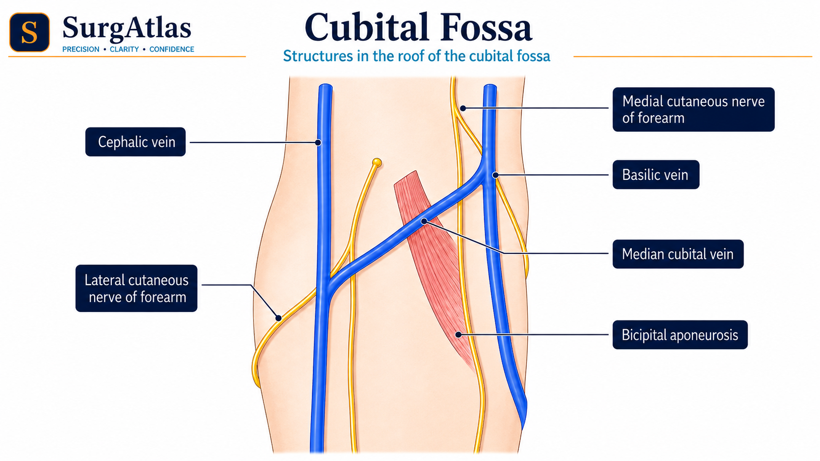
The median cubital vein is superficial to the bicipital aponeurosis.
Boundaries
| Part | Structure |
|---|---|
| Superior | Line joining medial and lateral epicondyles |
| Lateral | Brachioradialis |
| Medial | Pronator teres |
| Floor | Brachialis proximally and supinator distally |
| Roof | Skin, superficial fascia, deep fascia and bicipital aponeurosis |
Deep contents: lateral to medial
A useful central sequence is biceps Tendon, brachial Artery, median Nerve — TAN. If the radial nerve at the lateral margin is included, the expanded sequence becomes radial nerve, tendon, artery, nerve.
| Structure | Key relation |
|---|---|
| Radial nerve | At the lateral side, dividing into superficial and deep branches near the fossa. |
| Biceps tendon | Passes to the radial tuberosity. |
| Brachial artery | Usually divides into radial and ulnar arteries in or near the fossa. |
| Median nerve | Most medial major deep neurovascular structure. |
Why the roof matters
The median cubital vein and other superficial veins lie in superficial fascia. The bicipital aponeurosis fans medially from the biceps tendon and provides a protective layer over the brachial artery and median nerve.
Applied anatomy
- The brachial pulse is palpated medial to the biceps tendon.
- The fossa is an access site for venepuncture and blood-pressure assessment.
- Supracondylar injuries can threaten the brachial artery and median/anterior interosseous nerve structures.
- The radial nerve relationship explains why lateral elbow pathology can produce radial-nerve symptoms.
Active recall
Close the notes and answer these
Try each question from memory before revealing the answer. These public prompts are a small preview of the integrated retrieval system inside SurgAtlas.
01What is the lateral-to-medial TAN sequence?
Biceps tendon, brachial artery, median nerve.
02Which two muscles form the floor?
Brachialis and supinator.
03What separates the median cubital vein from the brachial artery?
The bicipital aponeurosis and deep fascia.
Sources & editorial basis
References & editorial basis
- SurgAtlas production chapter — Anatomy — Upper Limb I. Primary source for this public lesson. The teaching has been condensed from the corresponding production chapter without changing the underlying anatomical relationships.
- Last’s Anatomy: Regional and Applied. Reference for stable cubital-fossa relationships.
This lesson is derived from the corresponding SurgAtlas production teaching material. Where the source makes current management or guideline claims, the public lesson uses the cited contemporary guidance. It is written for education and examination preparation, not as patient-specific clinical advice.
Editorial details
Medical Doctor (MD) · MRCS Part A · Physician · Surgical Educator
SurgAtlas is an educational resource. For patient care, verify current national guidance, local antimicrobial and transfusion policies, specialty pathways and individual patient factors.
Continue in SurgAtlas
Turn reading into retrieval.
Use the full learning workspace for chapter progress, integrated questions, review scheduling and exam-focused study tools. Free accounts do not include recurring AI usage.