High-yield map
Key points before the detail
- 01
Roots are C5–T1; trunks are upper C5–6, middle C7 and lower C8–T1.
- 02
Anterior divisions mainly supply flexor compartments; posterior divisions unite into the posterior cord.
- 03
Cords are named lateral, medial and posterior relative to the second part of the axillary artery.
- 04
The median nerve is formed by lateral and medial roots, so it draws fibres from both lateral and medial cords.
Roots and trunks
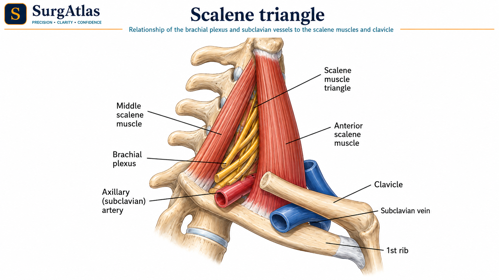
The ventral rami of C5–T1 emerge between anterior and middle scalene muscles. The subclavian artery passes through the same interscalene interval, while the subclavian vein lies anterior to anterior scalene. C5 and C6 unite as the upper trunk, C7 continues as the middle trunk and C8 with T1 forms the lower trunk.
- Dorsal scapular nerve commonly arises from C5.
- Long thoracic nerve receives C5–C7 contributions and descends on serratus anterior.
- The upper trunk gives the suprascapular nerve and nerve to subclavius.
Divisions and cords
| Cord | Formation | Major terminal branches |
|---|---|---|
| Lateral | Anterior divisions of upper and middle trunks | Musculocutaneous + lateral root of median |
| Medial | Anterior division of lower trunk | Ulnar + medial root of median |
| Posterior | All three posterior divisions | Axillary + radial |
Terminal branches
| Nerve | Principal compartment / function |
|---|---|
| Musculocutaneous | Anterior arm; elbow flexion and lateral forearm sensation |
| Axillary | Deltoid/teres minor; shoulder abduction and lateral shoulder sensation |
| Radial | Posterior arm/forearm extensors; dorsal hand sensory territory |
| Median | Most forearm flexors, thenar function and lateral palmar digits |
| Ulnar | Intrinsic hand emphasis and medial hand sensation |
Applied lesion patterns
- Upper-trunk injury preferentially affects C5–C6 functions such as shoulder abduction/external rotation and elbow flexion.
- Lower-trunk injury can markedly affect intrinsic hand function because C8–T1 fibres are prominent.
- A cord lesion produces a pattern involving several named terminal nerves; a terminal-nerve lesion is more anatomically restricted.
- Long thoracic nerve injury weakens serratus anterior and can produce medial scapular winging.
How to reconstruct the plexus in an exam
Write C5 to T1 vertically, combine C5–6, leave C7, combine C8–T1, split each trunk, then regroup anterior divisions into lateral and medial cords while all posterior divisions unite. Reconstructing the pattern is safer than relying on a memorised drawing without anatomical logic.
Active recall
Close the notes and answer these
Try each question from memory before revealing the answer. These public prompts are a small preview of the integrated retrieval system inside SurgAtlas.
01How is the posterior cord formed?
By union of the posterior divisions of all three trunks.
02Which cords contribute to the median nerve?
Both the lateral and medial cords, through lateral and medial roots.
03What is the relationship of the subclavian vein to anterior scalene?
It passes anterior to anterior scalene; the subclavian artery and brachial-plexus roots pass through the interscalene interval.
Sources & editorial basis
References & editorial basis
- SurgAtlas production chapter — Anatomy — Upper Limb I. Primary source for this public lesson. The teaching has been condensed from the corresponding production chapter without changing the underlying anatomical relationships.
- Gray’s Anatomy for Students. Reference for stable brachial-plexus organisation and relationships.
This lesson is derived from the corresponding SurgAtlas production teaching material. Where the source makes current management or guideline claims, the public lesson uses the cited contemporary guidance. It is written for education and examination preparation, not as patient-specific clinical advice.
Editorial details
Medical Doctor (MD) · MRCS Part A · Physician · Surgical Educator
SurgAtlas is an educational resource. For patient care, verify current national guidance, local antimicrobial and transfusion policies, specialty pathways and individual patient factors.
Continue in SurgAtlas
Turn reading into retrieval.
Use the full learning workspace for chapter progress, integrated questions, review scheduling and exam-focused study tools. Free accounts do not include recurring AI usage.