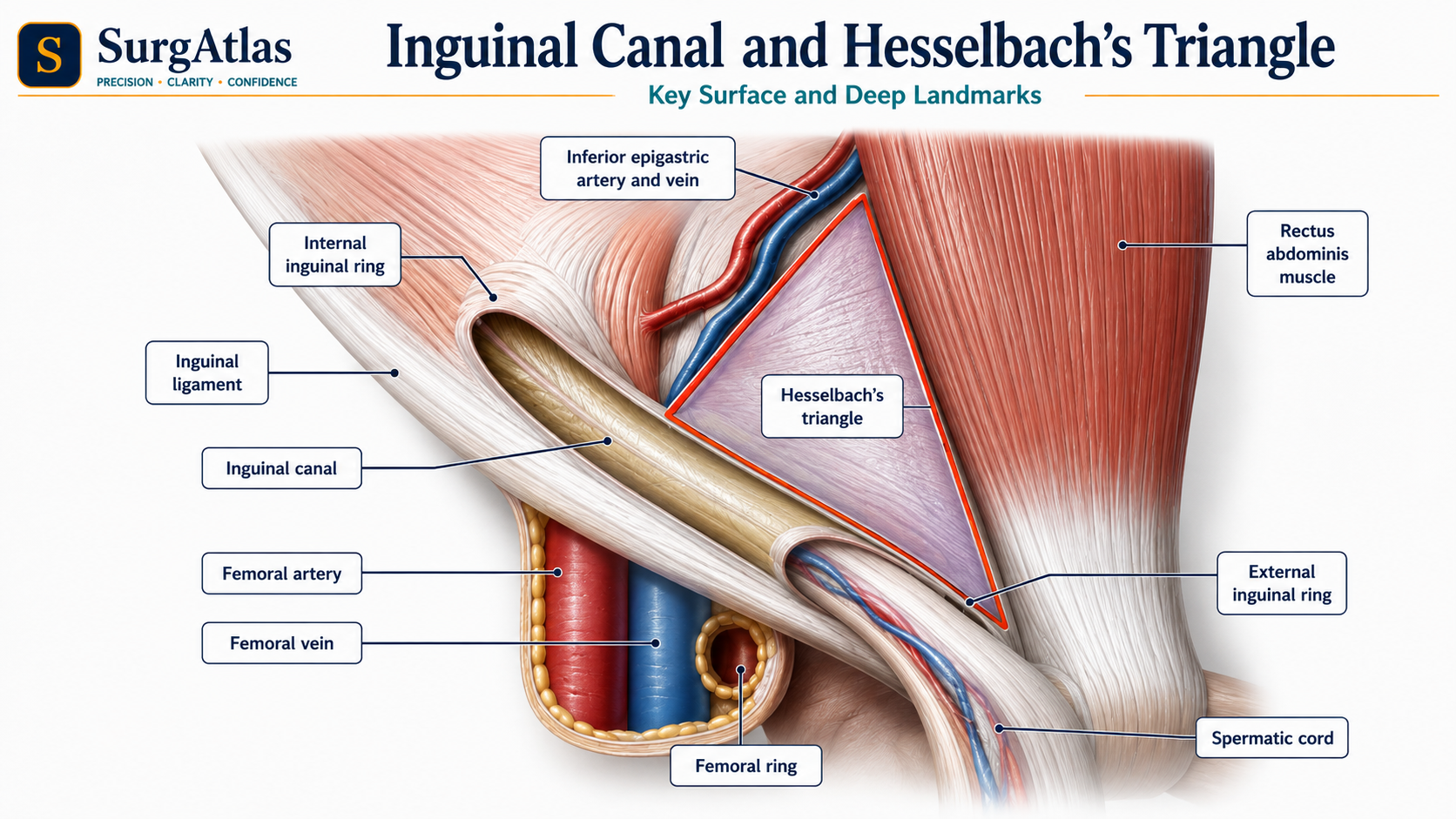
High-yield map
Key points before the detail
- 01
Deep ring: transversalis fascia, lateral to the inferior epigastric vessels.
- 02
Superficial ring: external oblique aponeurosis, just superolateral to the pubic tubercle.
- 03
The ilioinguinal nerve enters the canal separately and does not pass through the deep ring.
- 04
Indirect hernia = lateral to inferior epigastric vessels; direct hernia = medial.
- 05
The conjoint tendon is variable and should not be described as a universal robust structure.
1. Orientate the canal before memorising lists
The inguinal canal runs obliquely through the lower anterior abdominal wall from the deep inguinal ring to the superficial inguinal ring. In adults it is commonly described as about 4 cm long, although the exact length varies.
A useful mental model is to build the canal from deep to superficial. Start at the transversalis fascia, follow the spermatic cord or round ligament through the canal, then exit through the external oblique aponeurosis. Once that route is clear, the walls and hernia relationships become easier to reconstruct rather than memorise as isolated facts.
2. The deep and superficial rings
Do not confuse the midpoint of the inguinal ligament with the mid-inguinal point. The deep ring is described relative to the midpoint of the inguinal ligament. In examination answers, pair its surface position with its decisive vascular relationship: it is lateral to the inferior epigastric vessels.
| Structure | Anatomical position | Why it matters |
|---|---|---|
| Deep inguinal ring | Opening in transversalis fascia, about 1–1.5 cm above the midpoint of the inguinal ligament and lateral to the inferior epigastric vessels. | Entry point of an indirect inguinal hernia and the route through which the spermatic cord begins its passage. |
| Superficial inguinal ring | Triangular gap in the external oblique aponeurosis just superolateral to the pubic tubercle. | Exit of the canal into the superficial tissues; useful clinical surface relationship. |
3. The four walls of the inguinal canal
| Wall | Main component | Reinforcement or variation |
|---|---|---|
| Anterior | External oblique aponeurosis | Reinforced laterally by internal oblique. |
| Posterior | Transversalis fascia | Reinforced medially by a conjoint contribution when present. |
| Roof | Arching fibres of internal oblique and transversus abdominis | The muscular arch contributes to dynamic support of the canal. |
| Floor | Inguinal ligament | Reinforced medially by the lacunar ligament. |
4. Contents and spermatic-cord coverings
In the male, the canal transmits the spermatic cord plus the ilioinguinal nerve. In the female, it transmits the round ligament of the uterus plus the ilioinguinal nerve. The ilioinguinal nerve is an important exception: it enters the canal separately and does not pass through the deep ring.
The genital branch of the genitofemoral nerve travels within the spermatic cord and supplies the cremaster muscle as well as scrotal or labial skin. Testicular lymphatics travel with the cord but drain to para-aortic nodes, reflecting the embryological origin of the testis rather than its final scrotal position.
| Cord covering | Source layer |
|---|---|
| Internal spermatic fascia | Transversalis fascia at the deep ring |
| Cremasteric muscle and fascia | Internal oblique |
| External spermatic fascia | External oblique aponeurosis at the superficial ring |
5. Direct versus indirect inguinal hernia
The relationship of the hernia sac to the inferior epigastric vessels is the definitive anatomical distinction. Clinical examination can suggest the type, but it cannot reliably replace operative or imaging anatomy.
| Feature | Indirect inguinal hernia | Direct inguinal hernia |
|---|---|---|
| Inferior epigastric vessels | Lateral | Medial |
| Route | Enters the deep ring and may traverse the canal. | Pushes through the posterior wall in Hesselbach triangle. |
| Typical mechanism | Persistent processus vaginalis and/or widened deep ring. | Acquired weakness of the posterior wall. |
| Cord coverings | May acquire all cord coverings if complete. | Coverings vary with size and chronicity; does not enter through the deep ring. |
6. Hesselbach triangle
Notice that the inferior epigastric vessels serve two jobs in your mental map: they form the lateral boundary of Hesselbach triangle and they separate direct hernias medially from indirect hernias laterally.
- Medial boundary: lateral border of rectus abdominis.
- Lateral boundary: inferior epigastric vessels.
- Inferior boundary: inguinal ligament.
- Floor: transversalis fascia; direct hernias protrude through this region.
7. Clinical application and repair principles
A groin swelling should be assessed for site relative to the inguinal ligament and pubic tubercle, cough impulse, tenderness, reducibility and evidence of obstruction or strangulation. A painful irreducible swelling with systemic illness, skin change or bowel obstruction requires urgent surgical assessment rather than repeated forceful reduction.
For appropriately selected adults with minimally symptomatic inguinal hernia, watchful waiting can be a reasonable option with safety-netting. NICE recognises open repair, TEP and TAPP as treatment options; choice should consider anaesthetic suitability, whether the hernia is primary, recurrent or bilateral, the anatomy and the surgeon’s experience.
Active recall
Close the notes and answer these
Try each question from memory before revealing the answer. These public prompts are a small preview of the integrated retrieval system inside SurgAtlas.
01Where is the deep inguinal ring?
It is an opening in transversalis fascia about 1–1.5 cm above the midpoint of the inguinal ligament and lateral to the inferior epigastric vessels.
02State the four walls of the inguinal canal.
Anterior: external oblique aponeurosis, reinforced laterally by internal oblique. Posterior: transversalis fascia with a variable medial conjoint contribution. Roof: arching internal oblique and transversus abdominis. Floor: inguinal ligament, reinforced medially by lacunar ligament.
03Which nerve enters the canal without passing through the deep ring?
The ilioinguinal nerve.
04What is the definitive anatomical distinction between direct and indirect inguinal hernia?
Indirect hernias are lateral to the inferior epigastric vessels and enter the deep ring; direct hernias are medial and push through the posterior wall in Hesselbach triangle.
05State the boundaries of Hesselbach triangle.
Medial: lateral border of rectus abdominis. Lateral: inferior epigastric vessels. Inferior: inguinal ligament. The floor is transversalis fascia.
Sources & editorial basis
References & editorial basis
- SurgAtlas production source. Applied Surgical Anatomy — Abdomen I: inguinal canal, direct/indirect inguinal hernias and groin swelling assessment.
- NICE TA83. Laparoscopic surgery for inguinal hernia repair — open, TAPP and TEP decision factors and training requirements. Source ↗
- Evidence-Based Interventions. Repair of minimally symptomatic inguinal hernia — reviewed September 2024. Source ↗
This lesson is derived from the corresponding SurgAtlas production teaching material. Where the source makes current management or guideline claims, the public lesson uses the cited contemporary guidance. It is written for education and examination preparation, not as patient-specific clinical advice.
Editorial details
Medical Doctor (MD) · MRCS Part A · Physician · Surgical Educator
SurgAtlas is an educational resource. For patient care, verify current national guidance, local antimicrobial and transfusion policies, specialty pathways and individual patient factors.
Continue in SurgAtlas
Turn reading into retrieval.
Use the full learning workspace for chapter progress, integrated questions, review scheduling and exam-focused study tools. Free accounts do not include recurring AI usage.