
High-yield map
Key points before the detail
- 01
Name the layers from skin to peritoneum and know where the neurovascular plane lies.
- 02
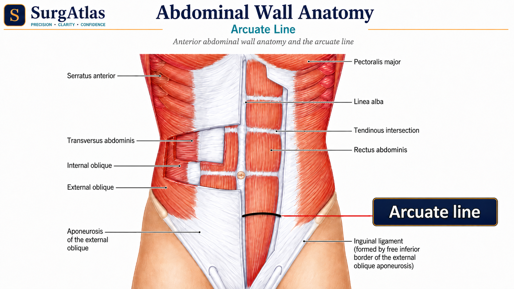
Above the arcuate line the posterior sheath is formed principally by posterior internal-oblique lamina and transversus aponeurosis; below it all aponeuroses pass anteriorly.
- 03
The arcuate line is variable and may be absent, so it should not be treated as a fixed universal landmark.
- 04
The inferior epigastric vessels arise from the external iliac vessels and are a major landmark for abdominal access and groin hernias.
Surface landmarks and planes
The abdomen can be described with four quadrants or nine regions. The nine-region scheme uses two midclavicular lines plus a subcostal plane near L3 and a transtubercular plane near L5. The transpyloric plane is classically drawn at L1 and is useful for orientation, but organ position varies with respiration, body habitus and posture.
Layers from superficial to deep
| Layer | High-yield relationship |
|---|---|
| Superficial fascia | Fatty Camper layer and more membranous Scarpa layer; Scarpa continues into the perineum but attaches to fascia lata below the inguinal ligament. |
| External oblique | Most superficial flat muscle; its aponeurosis contributes to the anterior rectus sheath and inguinal ligament. |
| Internal oblique | Splits around rectus above the arcuate line and contributes to both anterior and posterior sheaths. |
| Transversus abdominis | Deep flat muscle; its aponeurosis contributes posteriorly above the arcuate line and passes anteriorly below it. |
| Transversalis fascia | Continuous deep fascial layer behind the muscular wall. |
| Extraperitoneal tissue and parietal peritoneum | Final layers before the peritoneal cavity. |
The rectus sheath and arcuate line
Above the arcuate line, the external-oblique aponeurosis passes anterior to rectus. Internal oblique splits to contribute anterior and posterior laminae, while transversus abdominis contributes to the posterior sheath. Below the arcuate line, the aponeuroses of external oblique, internal oblique and transversus all pass anterior to rectus, leaving transversalis fascia directly behind the muscle.
Inferior epigastric vessels and the neurovascular plane
The inferior epigastric vessels arise from the external iliac vessels, enter the rectus sheath near the arcuate-line region and ascend on the posterior surface of rectus abdominis. They help distinguish direct from indirect inguinal hernias and are at risk during trocar or incision placement. Thoracoabdominal nerves travel mainly in the plane between internal oblique and transversus abdominis before supplying the abdominal wall.
- Deep epigastric vessels are not simply midline structures: identify them before lateral rectus or laparoscopic access.
- The internal-oblique/transversus plane explains why regional blocks can anaesthetise broad segments of the abdominal wall.
- Scarpa-fascia attachment to fascia lata helps explain why extravasated urine from an anterior urethral injury does not usually track freely into the thigh.
Surgical relationships to connect
- Midline laparotomy exploits the relatively avascular linea alba.
- Paramedian or muscle-splitting approaches require deliberate knowledge of rectus sheath, muscle and segmental nerves.
- Inferior epigastric vessels are an essential visual landmark in laparoscopic groin surgery.
- Weakness or denervation of abdominal-wall musculature can produce bulging without a true fascial defect.
Active recall
Close the notes and answer these
Try each question from memory before revealing the answer. These public prompts are a small preview of the integrated retrieval system inside SurgAtlas.
01What changes below the arcuate line?
All three flat-muscle aponeuroses pass anterior to rectus abdominis, leaving transversalis fascia directly posterior to the muscle.
02Where do the inferior epigastric vessels run in relation to rectus?
They arise from the external iliac vessels and ascend on the posterior surface of rectus within the sheath.
03Which plane carries the major segmental nerves of the anterolateral abdominal wall?
Mainly the plane between internal oblique and transversus abdominis.
Sources & editorial basis
References & editorial basis
- SurgAtlas production chapter — Anatomy — Abdomen I. Primary source for this public lesson. The teaching has been condensed from the corresponding production chapter without changing the underlying anatomical relationships.
- Last’s Anatomy: Regional and Applied. Standard applied-anatomy reference used for stable abdominal-wall relationships.
This lesson is derived from the corresponding SurgAtlas production teaching material. Where the source makes current management or guideline claims, the public lesson uses the cited contemporary guidance. It is written for education and examination preparation, not as patient-specific clinical advice.
Editorial details
Medical Doctor (MD) · MRCS Part A · Physician · Surgical Educator
SurgAtlas is an educational resource. For patient care, verify current national guidance, local antimicrobial and transfusion policies, specialty pathways and individual patient factors.
Continue in SurgAtlas
Turn reading into retrieval.
Use the full learning workspace for chapter progress, integrated questions, review scheduling and exam-focused study tools. Free accounts do not include recurring AI usage.