High-yield map
Key points before the detail
- 01
The rectum starts around S3 and lacks taeniae coli, appendices epiploicae and haustra.
- 02
Peritoneal covering decreases inferiorly: upper rectum anterior and lateral, middle anterior, lower rectum extraperitoneal.
- 03
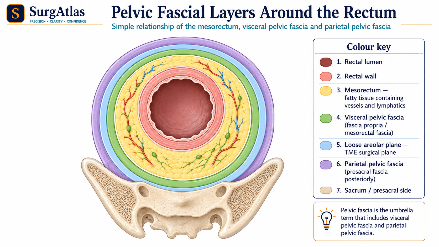
The mesorectum contains fat, vessels, lymphatics and nodes within visceral pelvic fascia.
- 04
TME follows the plane around the intact mesorectal fascia; leaving that plane can injure hypogastric and pelvic autonomic nerves.
Where the rectum begins and ends
The rectum begins near S3 as sigmoid features disappear and the longitudinal muscle layer becomes continuous. It follows the concavity of the sacrum and terminates at the anorectal junction, where puborectalis creates the anorectal angle.
Peritoneal relations
| Rectal level | Typical peritoneal covering |
|---|---|
| Upper third | Anterior and lateral surfaces |
| Middle third | Anterior surface |
| Lower third | No peritoneal covering |
What is the mesorectum?
The mesorectum is the fatty tissue surrounding the rectum that contains the superior rectal vessels, lymphatic channels and lymph nodes. It is enclosed by visceral pelvic fascia, commonly called the mesorectal fascia on imaging and in rectal-cancer surgery.
The TME plane
Total mesorectal excision aims to remove the rectum and mesorectal envelope intact by dissecting in the embryological fascial plane between the visceral mesorectal fascia and parietal pelvic fascia. The plane is relatively avascular when correctly entered.
- Staying on the mesorectal envelope supports a clear circumferential resection margin.
- Straying too laterally or posteriorly can increase bleeding and autonomic-nerve injury.
- The exact operative plane changes around anterior pelvic structures and must be adapted to sex, tumour location and previous treatment.
Pelvic autonomic nerves
The superior hypogastric plexus continues as paired hypogastric nerves, which join pelvic splanchnic parasympathetic fibres to form the inferior hypogastric plexuses. These fibres run close to the mesorectal and lateral pelvic planes. Injury can produce urinary and sexual dysfunction.
Active recall
Close the notes and answer these
Try each question from memory before revealing the answer. These public prompts are a small preview of the integrated retrieval system inside SurgAtlas.
01What is contained within the mesorectum?
Perirectal fat containing vessels, lymphatics and lymph nodes, enclosed by visceral pelvic/mesorectal fascia.
02What is the principle of the TME plane?
Dissect around the intact visceral mesorectal fascia in the relatively avascular plane between it and parietal pelvic fascia.
03Why does pelvic autonomic preservation matter?
Injury to hypogastric, pelvic splanchnic or inferior hypogastric plexus fibres can cause urinary and sexual dysfunction.
Sources & editorial basis
References & editorial basis
- SurgAtlas production chapter — Anatomy — Abdomen III. Primary source for this public lesson. The teaching has been condensed from the corresponding production chapter without changing the underlying anatomical relationships.
- Gray’s Anatomy for Students. Reference for stable rectal, fascial and autonomic pelvic anatomy.
This lesson is derived from the corresponding SurgAtlas production teaching material. Where the source makes current management or guideline claims, the public lesson uses the cited contemporary guidance. It is written for education and examination preparation, not as patient-specific clinical advice.
Editorial details
Medical Doctor (MD) · MRCS Part A · Physician · Surgical Educator
SurgAtlas is an educational resource. For patient care, verify current national guidance, local antimicrobial and transfusion policies, specialty pathways and individual patient factors.
Continue in SurgAtlas
Turn reading into retrieval.
Use the full learning workspace for chapter progress, integrated questions, review scheduling and exam-focused study tools. Free accounts do not include recurring AI usage.